
Assessing the Pupil: A Guide to Pupillary Light Reflex and Reactivity in Neuro Exams
As a professional, it’s crucial to thoroughly understand the pupillary light reflex and its significance in neuro exams. The pupillary light reflex is a crucial aspect of pupil assessment that can provide valuable insights into a patient’s neurological state.
This blog post is a comprehensive guide to understanding the pupillary light reflex and its role in neuro exams. By the end of this post, you will have a deeper understanding of the importance of this reflex in assessing a patient’s neurological health and be equipped with the knowledge necessary to effectively assess pupillary reactivity.
Understanding the Pupillary Light Reflex
The pupillary light reflex is a crucial component of neuro exams, as it reflects the health and functioning of the oculomotor, trochlear, and abducens nerves, as well as the integrity of the iris sphincter muscle and the optic nerve.
Physiology Behind Pupillary Light Reflex
When a bright light is shined into one eye, the iris sphincter muscle contracts, causing the pupil to constrict. This response occurs because the light stimulates the photosensitive cells in the retina, which send signals to the Edinger-Westphal nucleus in the midbrain. From there, the parasympathetic fibers of the oculomotor nerve activate the iris sphincter muscle, producing the pupillary light reflex.
Factors that Affect Pupillary Light Reflex
Various factors can influence the pupillary light reflex. For example, aging can cause a decrease in the speed and magnitude of the pupillary light reflex, while certain medications, such as anticholinergics, can interfere with the reflex. Conditions such as Parkinson’s disease, stroke, and brain tumors can affect the pupillary light reflex, so it is important to interpret the results of a pupillary light reflex test in the context of the patient’s overall health.
Importance of the Pupillary Light Reflex in Neuro Exams
The pupillary light reflex is an early indicator of neurological disorders and is crucial in differentiating between various conditions. It is an essential part of any comprehensive neuro exam. It plays a significant role in the differential diagnosis of neurological disorders.
Early Indicator of Neurological Disorders
The pupillary light reflex is critical for assessing neurological function and can detect issues early on. Abnormalities in pupil reactivity can indicate problems with the optic nerve or brainstem. They may be a sign of more significant neurological issues.
Differential Diagnosis
To arrive at an accurate diagnosis, it is crucial to differentiate between various conditions in a neuro exam. A pupillary light reflex is an excellent tool for differentiating between conditions such as neurological injury, infections, and other disorders that affect the central nervous system.
Monitoring Progress of Neuro Disorders
For patients diagnosed with a neurological disorder, a pupillary light reflex is a valuable tool for monitoring progress and determining the effectiveness of treatment. Changes in pupil reactivity can indicate a worsening condition or an improvement, providing critical information to medical professionals.
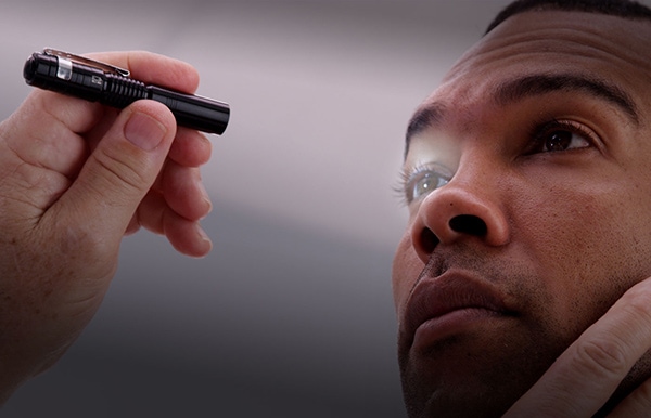
Steps for Conducting the Pupillary Light Reflex Test
As a healthcare professional, it’s important to understand the pupillary light reflex test and how to conduct it accurately. In this section, we’ll go over the preparation and equipment, the procedure for testing, and how to interpret the results.
Preparation and Equipment Needed
Before conducting the pupillary light reflex test, you’ll need to gather the equipment, including a penlight and a ruler or measuring tape. Consider having a bed or examination table and a quiet, well-lit environment for the patient to lie in.
Procedure for Testing Pupillary Light Reflex
Once your equipment is ready, begin by having the patient lie down or sit comfortably with their head facing forward. Next, use the penlight to shine a light into one of the patient’s eyes and observe the reaction of their pupil. Record the size of the pupil using the ruler or measuring tape, then repeat the process with the other eye.
How to Interpret Results?
The pupillary light reflex test results can assess the functioning of the patient’s optic nerve and the neural pathways involved in the pupillary response. A normally functioning pupillary light reflex will constrict the pupil when the light is shone into the eye and dilate when the light is removed. Any abnormalities in the pupillary response may indicate a neurological issue and should be further investigated using additional neurological tools, such as the NPi pupilometer.


